The X-ray detector performances
were assessed with 17.4 keV photons
at the METROLOGY beamline. The overall
gain value was measured to be 0.005
ADU/X-ray, therefore in accordance
with the absorption of the 20µm YAG:Ce
scintillator and the objective collection
efficiency evaluated [3]. The X-ray detector
spatial resolution was deduced from
the modulation transfer function (MTF)
calculated by Fourier transform of the Line
Spread Function obtained with a classical
knife edge method [4]. The measurement
is in good agreement with the analytical
model with 10% modulation transfer
contrast obtained around 61 cy/mm,
corresponding to a detail object of 8 µm.
Finally, the Detective Quantum Efficiency
(DQE) was infered from the Noise power
spectrum measurement. The calculated
DQE (0 cy/mm) is only 2% and this low
value is due to the poor X-ray absorption
of the scintillator and the limited light
collection of the optics.
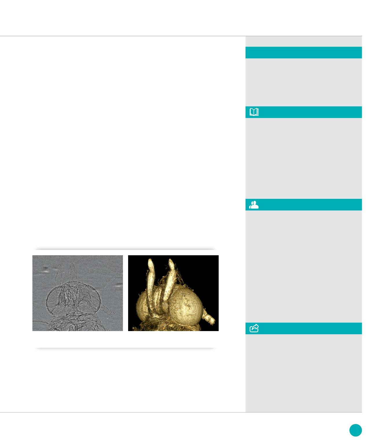
The first phase-contrast tomography
reconstruction of the head of a small
insect (food moth) has been carried
out on the METROLOGY beamline and
demonstrates clearly the performance
of this X-ray imaging detector. For this
tomographic acquisition, 2000 images
over 180° have been taken.
The tomograms were reconstructed,
see in figure
➋
.
These results, in particular the spatial
resolution, shows that the expertise
gained with the development and
characterization of many beam viewers
or X-ray cameras enables SOLEIL to
design, build and qualify to completeness
more complex imaging systems. The
skills of the Detector and Design and
Engineering groups, the capabilities of the
Mechanical workshop and the availability
of the X-ray beam and of a versatile end
station on the METROLOGY beamline allow
to design instruments exactly tailored
to the experimental requirements
of the imaging applications of SOLEIL.
The detector shall be used soon to
produce a fast (a few minutes only) large
tomography reconstruction from a few
thousand images, taking advantage
of a high flux white beam and of the high
frame rate of the camera [5]. Furthermore,
other detectors are currently designed
in house for the tomography beamlines
of SOLEIL.
Detector & Conception
Engineering Groups
ASSOCIATED PUBLICATION
MTF, NPS and DQE characterization of an
in-house developed X-ray imaging detector
for synchrotron based micro-tomography
K. Desjardins*, M. Bordessoule, S. Hustache,
C. Menneglier, A. Carcy, S. Bonnin
and K. Medjoubi
IWORID 2013 Proceeding
* Synchrotron SOLEIL, l’Orme des Merisiers,
St Aubin BP48, 91192 Gif sur Yvette Cedex,
France
CORRESPONDING AUTHOR
➋
Image of the head of a moth (
a
) In-line phase contrast. (
b
) Volume rendering of the Edge Enhancement
Computed Tomography.
REFERENCES
[1] P.-A. Douissard et al. 2012 JINST 7 P09016
[2] PCO,
[3] Lui et al, Med. Phys. 21 (1994) (7):1193
[4] International Standard ISO 12233:2000(E)
[5] K. Medjoubi et al. J. Synchrotron Rad.
20(2) (2013) 293
(
a
)
(
b
)
117
SOLEIL
HIGHLIGHTS
2013


