An in-house developed
X-ray imaging detector
for synchrotron based
micro-tomography
Hard X-ray micro-tomography is a powerful
tool to reveal the internal structure
of thick objects in a non-destructive
manner. The unique characteristics of
synchrotron radiation, such as high flux,
monochromaticity, and partial coherence
facilitate sensitive fast tomography in
absorption and phase contrast modes, down
to micrometer spatial resolution. For these
imaging techniques, the performances
of the detector are particularly important [1].
The Detectors and Design & Engineering
groups at SOLEIL, in collaboration with
Paris Sud Orsay University, have designed,
developed, manufactured and assembled
a fast, sensitive and high resolution indirect
imaging X-ray detector with the goal of
carrying out preliminary micro-tomography
test experiments on SOLEIL beamlines, and
to be used by students for practical classes.
We present here the performances of the
X-ray detector deduced from measurements
on the METROLOGY beamline and we
demonstrate the feasibility of phase contrast
tomographic reconstruction with a test
sample.
The detector is based on a thin single
crystal scintillator screen YAG:Ce (20μm
freestanding) coupled with a low distortion
objective (QIOPTIC Inspec.xL 105mm f/4 –
G3) allowing to magnify by three the image
onto a new generation CMOS scientific
sensor (2560 x 2160 pixels)
of the low noise and high speed (100Hz)
PCO edge camera [2]. The field of view
is 4.7 mm x 5.5 mm with pixels of 2.3µm.
A 45° tilted mirror, inserted between
the scintillator and the objective,
reflects the visible light coming from
the scintillator out of the X-rays axis,
to prevent any radiation damage on
the sensor. The characteristics of these
components were beforehand measured in
laboratory with visible light and, especially,
the Numerical Aperture (NA)
of the objective and the PCO Edge camera
properties (dark noise, saturation capacity
and electronic gain). The measurement
results are summarized in Table 1.
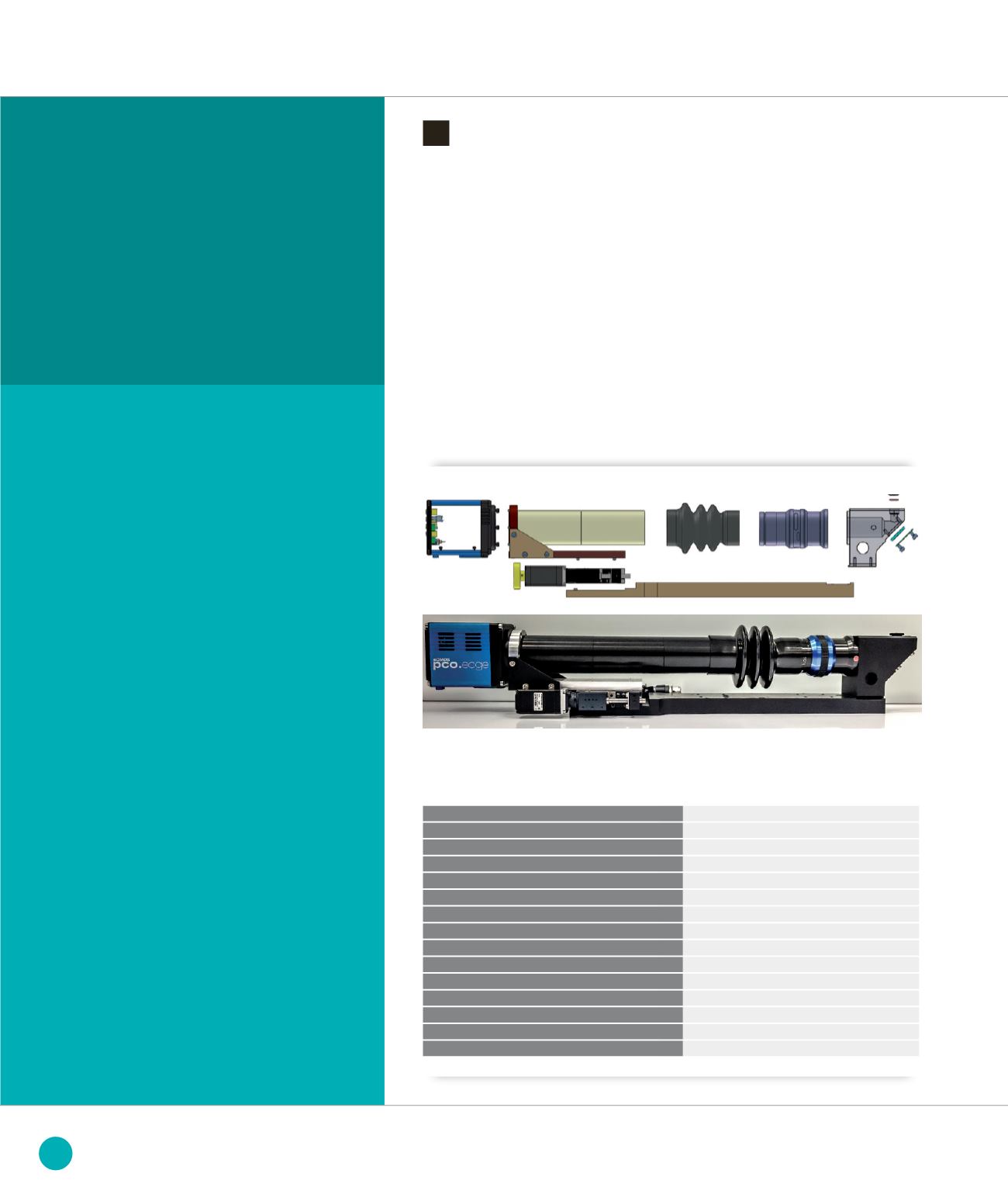
A mechanical assembly was designed
in-house, with a motorized translation
to focus the optics onto the scintillator
screen. A particular attention was given
to the parallelism between of the different
plans: sensor, mirror and scintillator
screen. Figure
➊
gives a detailed 3D view
of the mechanical concept and a picture
of the X-ray detector.
➊
X-ray tomography detector (
a
) Exploded 3D view of mechanical design (
b
) Final device photography
(
a
)
(
b
)
Camera
Scientific CMOS - PCO Edge
Frame rate
Max. 100 fps
Pixel format
2560 x 2160 pixel
Pixel size
6.5 µm
Analog-to-digital resolution
16 bits
Gain e− / analog-to-digital unit
0.545 e-/ADU
Saturation capacity
29155 e
-
Dark current
2-6 e
-
/pixel/s
Temporal dark noise
1.5 e
-
Objective
Qioptiq inspec.xL 105 mm f/4, M = 2.86
Numerical aperture (NA)
0.125
Working distance
81.3 mm
Scintillator
YAG:Ce
∅
9mm x 20.2 µm (+/-0.2µm)
Image size
5.55 mm x 4.17 mm
Pixel Image size
2.3 µm
TABLE 1: X-RAY DETECTOR CHARACTERISTICS
MODELING, METHODOLOGY AND INSTRUMENTATION
116
SOLEIL
HIGHLIGHTS
2013


