Contact : Marie-Joëlle ANTOINE
Tel : + 33(0)1 69 35 90 30 ou + 33(0)1 69 35 91 40
Mail : industrie@synchrotron-soleil.fr
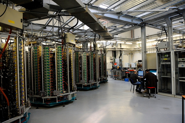
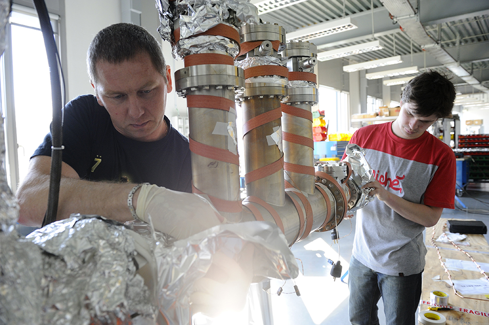
SOLEIL est une plateforme de caractérisation et d’étude des matériaux (vivants, inertes, biomatériaux, surfaces, plasma, liquides, matrices complexes...) qui utilise les techniques synchrotron telles que les spectroscopies et nano ou micro-imageries (X, UV, IR, ou XPS), la diffraction des rayons X (monocristaux, surfaces, poudres, biocristallographie...), les techniques de diffusion (SAXS, GISAXS...) ou encore la micro et nano-tomographie.
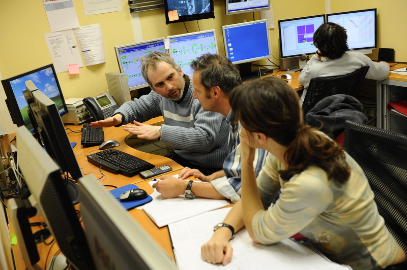
SOLEIL propose des techniques de pointe et son expertise en analyse de matériaux, au sein de 29 laboratoires d'analyse spécialisés et 29 équipes dédiées (scientifiques réalisant leur propre recherche et accueillant chaque jour de nombreux utilisateurs issus de disciplines scientifiques et secteurs variés).
Pour en savoir plus sur les services sur mesure proposés aux entreprises ou laboratoires souhaitant accéder à des moyens de hautes performances pour analyser, caractériser, contrôler ou observer jusqu'à l'échelle du nanomètre vos matériaux, n'hésitez pas à nous contacter.