IPANEMA
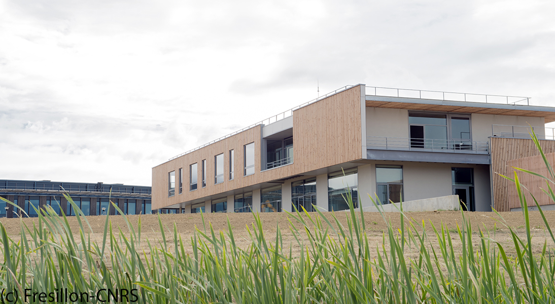
Institut photonique d'analyse non-destructive européen des matériaux anciens, IPANEMA est une plateforme de recherche, unique au niveau international, dédiée à l'étude des matériaux anciens à SOLEIL.
NanoimagesX
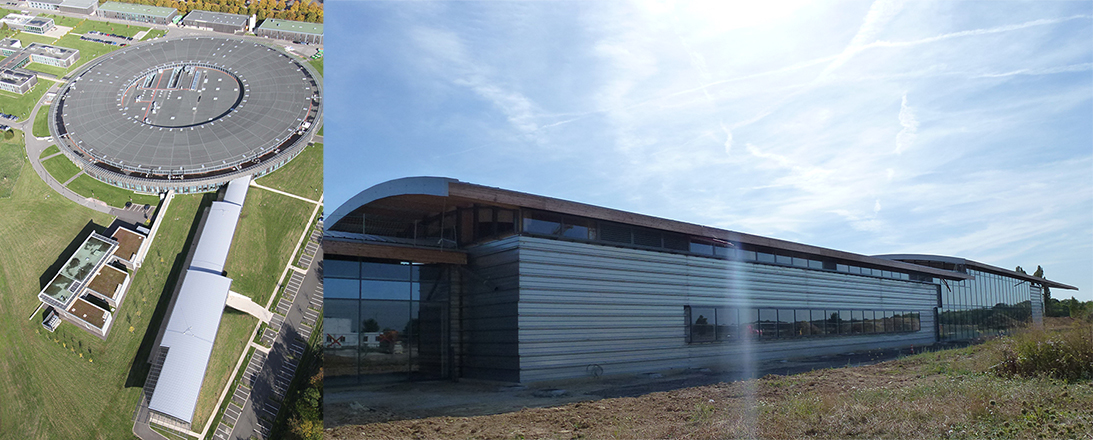
NanoimagesX est un Investissement d'Avenir (deuxième appel à projet EQUIPEX) au synchrotron SOLEIL, destiné aux utilisateurs académiques et industriels des domaines des Sciences de la vie et des matériaux.
MICASOL
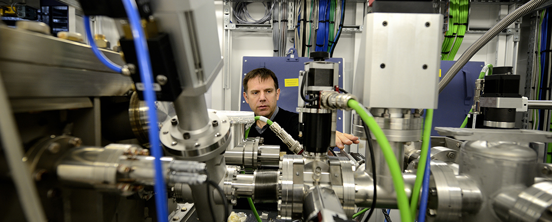
MICASOL est une plateforme ouverte aux industriels de la Région Grand Est de tous secteurs, intéressés par les nanotechnologies et la caractérisation des matériaux, de l’échelle moléculaire à l'échelle macroscopique.
Cosmetomics
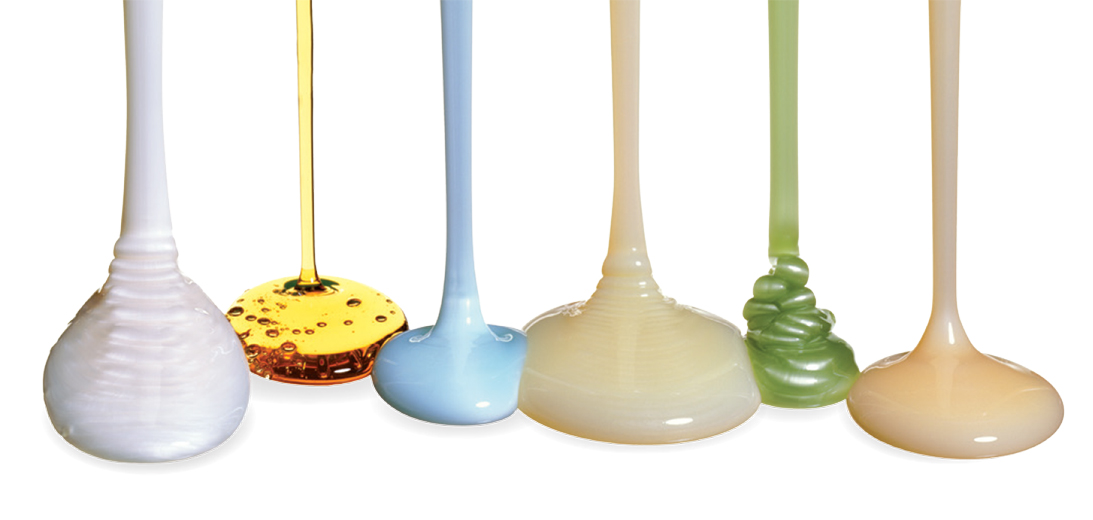 Cosmetomics est une plateforme mutualisée d’innovation (PFMI) à destination des entreprises françaises du secteur de la cosmétique avec comme objectif la caractérisation et à la mesure de l’efficacité et de l’innocuité de leurs produits.