DEIMOS, Dichroism Experimental Installation for Magneto-Optical Spectroscopy, est une ligne de lumière dédiée à l’étude des propriétés magnétiques et électroniques grâce au rayonnement synchrotron polarisé dans le domaine des rayons-X de basse énergie.
 L’utilisation du rayonnement synchrotron dans le domaine du magnétisme repose sur le développement des sources de rayons-X polarisés circulairement et linéairement. L’avantage des techniques de dichroïsme* magnétiques utilisées sur DEIMOS, comparé à d’autres techniques de magnétométrie, réside dans la capacité à mesurer la structure électronique et magnétique de chaque élément chimique indépendamment. De plus, de par la nature dipolaire de l’Hamiltonian d’interaction, il est possible pour des seuils d’absorption spécifiques de déterminer les moments magnétiques d’orbite et de spin. Par ailleurs, grâce à une limite de détection extrêmement faible, ces mesures peuvent être réalisées sur des quantités infimes de quelques atomes ou molécules. Cet ensemble de propriétés rend ces techniques parfaitement adaptées aux études scientifiques liées au développement des matériaux innovants qui nécessitent une optimisation des propriétés physiques et chimiques. Dans ce cadre les techniques de dichroïsme peuvent apporter des réponses spécifiques sur les mécanismes de couplages magnétiques ou de transport pour ne citer que quelques exemples.
*différence d’absorption entre rayons-X polarisés circulaire droit et gauche ou linéaire vertical et horizontal en présence d’un champ magnétique (XAS/XMCD, XMLD).

L'équipe


Offres d'emploi & de stage
Consultez les offres d'emploi de SOLEIL.
Current OPEN POSITION (deadline 11th of August 2023)
PhD position between DEIMOS - Synchrotron SOLEIL (Paris) and the University of Florence (Firenze Italy) on "LARGE SCALE FACILITIES BASED ULTRA-LOW TEMPERATURE MAGNETIC INVESTIGATION OF MOLECULAR SYSTEMS ASSEMBLED ON SURFACES".
Open position in collaboration between DEIMOS beamline at the SOLEIL Synchrotron and the Laboratory of Molecular Magnetism of the University of Florence (ITALY) for a specific project focused on the study at ultralow temperature (bellow 1K) of magnetic molecules. It is a 36 months PhD scholarship and it foresee 24 months stay in Firenze + 6 months stay at the SOLEIL Synchrotron + 6 months stay in another research institute to be defined with an agreement between the supervisor and the PhD student.
For further information contact: Prof. Matteo Mannini matteo.mannini@unifi.it
To apply: https://www.unifi.it/p12402.html
Données techniques
350 – 2500 eV
E/ΔE entre 6000 et 10000 sur toute la gamme d'énergie.
1er - APPLE-II HU52 : Polarisations circulaires gauche – droite, linéaires verticales et horizontales et polarisations tiltées.
2ème- EMPHU65 : possibilité de switcher entre polarisations circulaires droite et gauche à une fréquence de 5 Hz.
~ 2x10¹⁵ phot/s/0.1%bw @ 750eV
Double miroir (2 miroirs avec 2 angles d'incidence, permettant une bonne réjection des harmoniques sur toute la gamme d'énergie) ;
PGM avec réseaux VGD fonctionnant en mode Petersen + réseaux multicouches pour la gamme d'énergie "élevée" (>1500eV) ;
Système Wolter à post-focalisation.
La conception de l'optique est destinée à donner une grande stabilité et une grande pureté spectrale.
~80x80 µm² et ~800x800 µm2
~ 6x10¹² phot/s/0.1%bw @ 750eV
CroMag : TEY (Total Electron Yield), TFY (Total Fluorescence Yield - diode UHV) et transmission.
MagneTwo : TEY (Total Electron Yield), TFY (Total Fluorescence Yield - diode UHV ou détecteur SDD).
Thématiques scientifiques
| SURFACE ET INTERFACE MAGNÉTISME, MATÉRIAUX POUR la spintronique |
Propriétés magnétiques des structures de petite dimension - effets de taille ; corrélation entre les propriétés magnétiques, la morphologie et la structure - effets magnétoélastiques, contributions à l'anisotropie magnétique ; moments magnétiques et anisotropie des atomes isolés - diffusion superficielle par effet tunnel à très basse température ; interfaces ferromagnétiques-antiferromagnétiques - origine du couplage d'échange ; jonctions magnétiques à effet tunnel ; etc. |
|---|---|
| Aimants moléculaires, Films de Langmuir-Blodgett, Matériaux magnétiques hybrides |
Propriétés magnétiques et électroniques des aimants moléculaires purs (dichroïsme de la petite polarisation magnétique des groupes NO, avec sélectivité du site) ; composés organo-métalliques et assemblages supramoléculaires - grande variété de structures magnétiques accordant les propriétés chimiques, structures et mécanismes magnétiques "exotiques" ; composés lamellaires - ferrimagnétisme, frustration magnétique ; molécules polynucléaires aux propriétés magnétiques monodisperses (moments et anisotropie) - électronique moléculaire, q-bits. |
| Superparamagnétique nanoparticules, Sciences de la Terre, Paléomagnétisme |
Propriétés magnétiques de particules de magnétite (Fe3O4), de maghémite (γ-Fe2O3), d'hématite (α-Fe2O3), de pyrrhotite (Fe1-XS) ou de greigite (Fe3S4) diversement synthétisées - inclinaison de la surface magnétique, désordre chimique et magnétique. |
Full description of the beamline can be found in the following reference : Ohresser, P., Otero, E., Choueikani, F., Chen, K., Stanescu, S., Deschamps, F., Moreno, T., Polack, F., Lagarde, B., Daguerre, J.P., Marteau, F., Scheurer, F., Joly, L., Kappler, J.P., Muller, B., Bunau, O., Sainctavit, P. "DEIMOS: A beamline dedicated to dichroism measurements in the 350–2500 eV energy range" Review of Scientific Instruments, 85(1): art.n° 013106. (2014).

Sources
The photon source is constituted by two undulators : the HU-52, a 1.65 m long Apple-II helical undulator with a period of 52.4 mm, providing fully polarized light over the full energy range using the first, second and third harmonics. With circularly polarized light (left and right) the first harmonic covers the range ~260–1300 eV while horizontal and vertical linearly polarized light covers the range from 350 eV to 1150 eV.
The second undulator is an hybrid electromagnet/permanent magnet helical undulator (EMPHU) with a period of 65.0 mm optimized for a fast switching (5 Hz) of the circular polarized light, with no access to the pure linear horizontal polarization.

Apple-II undulator (left) ; EMPHU undulator (right)
The buffer chamber and DiagOn
The buffer chamber and the DiagOn (for Diagnostic Onduleur) are located in the radiation-protection hutch.
The pink beam emitted by the undulators can be visualized and characterized using the DiagOn device; diagnostic tool intended to precisely define the emission axis of the undulator [1].
The first optics chamber
Also located in the radiation-protection hutch, the first optics chamber (a.k.a. M1 chamber) serves basically to remove the unwanted beams : higher harmonic rejection, heat load, Bremsstrahlung…
It contains 3 mirrors that are water cooled : first a plane mirror (Pt coated SiC-CVD), that dissipates a large part of the 160W in-coming power, followed by two toroidal mirrors (Pt coated Si and Rh coated Si) to choose from which focus the beam into the monochromator.

Monochromator
The monochromator is equipped with two plane gratings which are respectively a variable groove depth (VGD) grating with 1600 lines/mm, and a Mo2C/B4C alternate multilayer (AML) grating with 2400 lines/mm. The VGD grating is optimized for the energy range 240-1500 eV while the AML grating is optimized for the energy domain 1000-2500 eV.
With the VGD grating the monochromator is usually operated in the Petersen geometry with a fixed c factor that can be choose to either maximize the flux with a poor resolution (large value) or minimize the flux with an improve resolution (small value down to 0.04). The energy resolution is further defined by the opening of the exit slits located 4m downstream of the main UHV vessel. The ultimate resolving power (exit slits closed) is around 30000 for a factor c of 0.2.

Refocussing (Wolter) chamber
The last optical stage is a simplified Wolter mirror combination associating a spherical mirror and a toroidal mirror. The choice between 2 spherical mirrors and 2 toroidal mirrors allows for 2 spot sizes ~80×80 mm2 and ~800×800 mm2 for the 2 working points (associate to the 2 workstations).

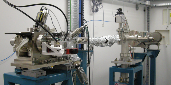
End-stations
Two end-stations are available on DEIMOS :
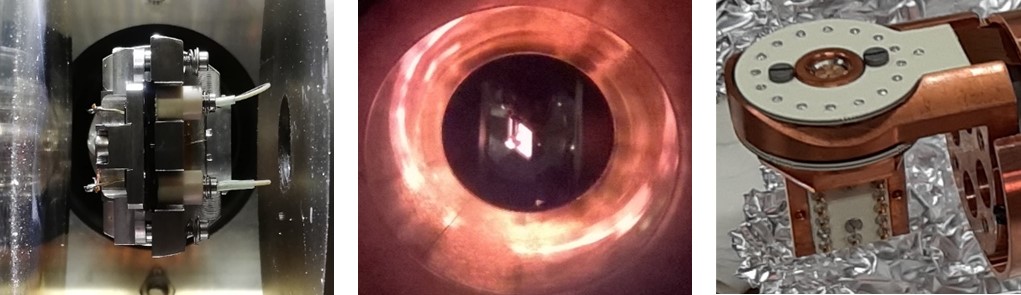
- A superconducting magnet (CroMag) providing ±7 T along the x-rays beam or ±2 T perpendicular to the beam. A variable temperature insert (V2TI) has been developed for temperatures in the 1.5-350 K range. A new dilution insert is in development to go in the ultra low temperature range and is intended to cover the 0.2-350 K temperature range [2].
- A 2T electromagnet (MagneTwo) allowing for a fast flipping of the magnetic field up to 1Hz. This end-station host several dedicated inserts: a high temperature insert (20-1000 K), an insert with multiple electrical connections (V²TI) (allowing for instance operando studies by applying voltages on a sample) [3] and a microfluidic insert for the studies of liquids.

1- CroMag ; 2- MagneTwo ; 3- STM

Two endstations : CroMag (left) ; MagneTwo (right)
You will find below the informations concerning the endstations :

Here are three different types of inserts :
Liquid cell at the centre of the magnet (left), 1000K under soft X-rays beam (centre), T2VI insert (right).
[1] T. Moreno, E. Otero, P. Ohresser, "In situ characterization of undulator magnetic fields", Journal of Synchrotron Radiation 19(2): 179-184 (2012)
[2] Joly, L., Muller, B., Sternitzky, E., Faullumel, J. G., Boulard, A., Otero, E., Choueikani, F., Kappler, J. P., Studniarek, M., Bowen, M. and Ohresser, P., "Versatile variable temperature insert at the DEIMOS beamline for in situ electrical transport measurements", Journal of Synchrotron Radiation, 2016, 23 (3): 652-657.
[3] J.-P. Kappler, E. Otero, W. Li, L. Joly, G. Schmerber, B. Muller, F. Scheurer, F. Leduc, B. Gobaut, L. Poggini, G. Serrano, F. Choueikani, E. Lhotel, A. Cornia, R. Sessoli, M. Mannini, M.-A. Arrio, Ph. Sainctavit, and P. Ohresser, "Ultralow-temperature device dedicated to soft X-ray magnetic circular dichroism experiments", Journal of Synchrotron Radiation, 25(6): 1727-1735. (2018)
Sample preparation chambers
Connected to the end-station, there are several equipment allowing for the introduction and the preparation of the samples :
- A fast load-lock is used to transfer sample in the UHV environment common to both endstations. The load-lock can be connected to a glove box (6) allowing a transfer through inert atmosphere. Beside, the use of a UHV suit case is also possible to transport sample from a nearby laboratory to our set-up. Contact your local contact if you whish to use DEIMOS’s suitcase.
- The "MBE" chamber is hosting several UHV equipment for in situ sample characterization and preparation:
- Sample preparation :
- Ion sputtering,
- Omniax manipulator with a temperature range of 150-700K,
- Evaporation (also possible on the small side chamber Raoul-petite) :
- 2 Knudsen cells,
- 2 effusion cells for organics (5),
- 3 electrons beam evaporators (Omicron EFM-3 and EFM-3T) (3),
- Quartz microbalance,
- Sample characterization :
- Auger analyser (4),
- LEED (Low Energy Electron Diffraction) (2),
- Variable temperature STM (50-500K) (1).
- Sample preparation :

Porte échantillons
Nous pouvons analyser plusieurs types d’échantillons : massifs, dépôts sur substrats ou sur monocristaux, transparent ou poudre. Il est également possible d'étudier des fluides grâce à l'insert microfluidique. Les échantillons peuvent être préparés à votre institut ou directement sur la ligne grace à notre ![]() MBE (1016.06 Ko)(pdf).
MBE (1016.06 Ko)(pdf).
CroMag:
Nous utilisons principalement 2 types de porte-échantillons dans le CroMag: les blocs de cuivre et les plaquettes de type Omicron.
Les échantillons peuvent être fixés directement sur les blocs de cuivre porte-échantillons (![]() cliquez ici pour schéma d'un porte-échantillon (59.65 Ko)) grace à des masques en cuivre (voire figure a-c) faits à façon; taille maximale des échantillons 9x25mm2.
cliquez ici pour schéma d'un porte-échantillon (59.65 Ko)) grace à des masques en cuivre (voire figure a-c) faits à façon; taille maximale des échantillons 9x25mm2.

Figure a : Porte échantillons avec plusieurs types de masques en cuivre; taille et forme à façon.
Figure b : Porte échantillons pour transmission.
Figure c : Porte échantillon avec 3 échantillons.
Les échantillons peuvent également être montés sur des ![]() plaquettes de type Omicron (62.99 Ko)(pdf) disponibles sur DEIMOS, et peuvent être maintenus par des fils, des pattes soudés, ou des vis. Il est également possible d’utiliser nos plaquettes surélevées ; taille maximale des échantillons 10mm de diamètre (figure d).
plaquettes de type Omicron (62.99 Ko)(pdf) disponibles sur DEIMOS, et peuvent être maintenus par des fils, des pattes soudés, ou des vis. Il est également possible d’utiliser nos plaquettes surélevées ; taille maximale des échantillons 10mm de diamètre (figure d).
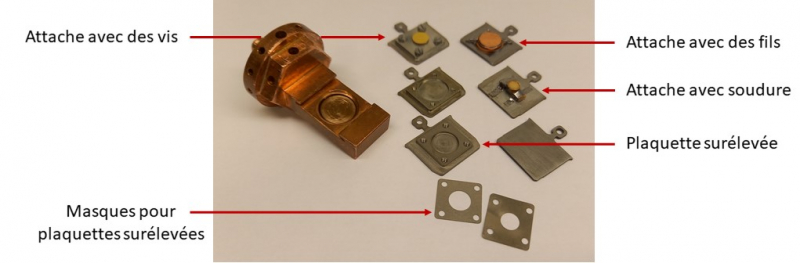
Figure d : porte échantillon pour plaquette de type Omicron.
Nous disposons également d’un porte échantillon tournant (figure e); taille maximale de l’échantillon 9x9mm2.
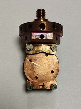
Figure e : porte échantillon tournant.
MagneTwo:
Dans MagneTwo, pour les mesures avec l'insert MilleK, les échantillons doivent être montés sur des plaquettes de type Omicron, pour l'insert V²TI nécessitant des contacts électriques les échantillons sont montés sur des puces électroniques enfichées sur un bloc de cuivre dédié (voir figure g) et pour l'insert microfluidique nous utilisons des puces microfluidiques (voir figure i).

Figure f : exemple d'échantillons montés sur deux puces électriques pour V2TI.
Figure g : puces électriques avec leur bloc de cuivre dédié.
Figure h : puce électrique en place sur l'insert V2TI.

Figure i : chip microfluidique.
Chargement des échantillons
Pour insérer les échantillons préparés ex-situ, l’insertion peut se faire via le « fast load lock » du SAS (voir photo ci-dessous), ou à travers la boite à gants (sous atmosphère inerte). Il est également possible d'utiliser une valise UHV que l'on peut connecter au SAS via une chambre de pompage intermédiaire (voir photos ci-dessous).


Valise UHV de 121,5cm.
Le parking de notre valise UHV peut loger jusqu’à 6 plaquettes de type Omicron (limité à 3 échantillons épais);

Contactez votre local contact pour plus d’informations concernant l’utilisation de la valise UHV de DEIMOS; clicker ici pour les informations fournisseur.
Contrôle et acquisition des données
On DEIMOS the interface to control the beamline is using scripting based on the PyTango biding [1]. With a small list of commands, it is possible to launch "macro" consisting of a series of actions/commands (launching scans, moving actuators, …) written in Python in a text file easy to edit and modify.
Acquisition usually mostly consists in cumulating absorption spectra (performed at dedicated sample conditions and polarization) and magnetization curves.
There is 2 ways to record absorption spectra and the resulting dichroic spectra:
- turboscan: the turboscan is a continuous scan (or scan-on-the-fly) where the 2 rotations of the gratings and mirrors (both in the monochromators) are moved to cover the energy range of interest while at the same time the energy (gap) and polarization (phase) of the undulator is enslaved to the monochromator energy trajectory [2]. During these displacements the sensors, and actuators positions are synchronously recorded. This method allows for shorter acquisition time (no dead-times) and smoother data acquisition because there is only one single displacement. A typical turboscan (80eV wide) is obtained in 2-3 min. For a dichroic spectrum one needs at least 2 XAS but depending on the statistic and to cancel instrumental artefact it is usually mandatory to record many pairs.
- flipscan: the flipscan is a step-by-step scan where the light polarization (CR / CL, for circular right / left) (using the undulator EMPHU) or the magnetization (+H / -H) (2T electromagnet) is flipped at each point in energy. The step-by-step flipscan is typically 30-40 min long but is very powerful to resolve very small XMCD signals.
Similarly, there is 2 ways to measure magnetization curves:
- turbohyst: the sensors are synchronously recorded while the magnetic field is performing the desired trajectory in magnetic field. A complete magnetization curve is obtained by recording 2 turbohyst for the polarizations CR and CL with the energy set at the maximum of the XMCD signal. The total time requested is typically around 45 min (depends highly in the trajectory in field).
- fliphyst: the fliphyst is a step-by-step scan where the light polarization (CR / CL) (using the undulator EMPHU) is flipped at each point in magnetic field. The step-by-step fliphyst takes more time compared to the turbohyst but allows to resolve signal that are not easily measured with the later.
[1] PyTango documentation website.
[2] Joly, L., Otero, E., Choueikani, F., Marteau, F., Chapuis, L., & Ohresser, P., "Fast continuous energy scan with dynamic coupling of the monochromator and undulator at the DEIMOS beamline", Journal of Synchrotron Radiation, 2014, 21(3): 502–506
Contact
Philippe OHRESSER, Responsable de la ligne : 01 69 35 96 82, philippe.ohresser@synchrotron-soleil.fr
Numéro de la ligne DEIMOS : 01 69 35 81 51 ; 01 69 35 81 52
Adresse pour envoi de matériel :
Synchrotron SOLEIL
L'Orme des Merisiers
Departementale 128
91190 SAINT-AUBIN
FRANCE