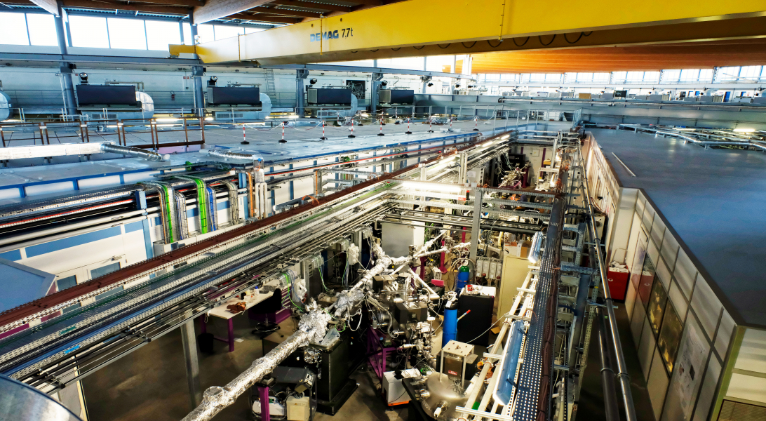
RESEARCH ON OUR SITE
 
 
Beamlines
Research Contents > Beamlines

The experimental facilities of a synchrotron beam center are called “beamlines.” These consist of a group of three successive cabins where the beam is captured, selected, focused, and directed toward the samples being studied. Each beamline is specialized by type of energy (at SOLEIL, from infrared to X-ray). Each line can be used for one or more analytical techniques: chemistry, physics, materials, biology, medicine, environment, astrophysics, heritage—each line can be used in one or more areas of application.

Beamlines per beam spot size Beamlines per photon energy range
Beamlines per analysis method  


Beamlines per beam spot size
Beam spot size List of Beamlines
<10µm ANTARES, LUCIA, SMIS, Microfocus, NANOSCOPIUM, GALAXIES
From 10 to 100µm DEIMOS, PLEIADES, TEMPO, PROXIMA 2, PSICHÉ, GALAXIES, MARS
From 100 to 300µm DISCO, MARS, PLEIADES, PROXIMA 1, SAMBA, DESIRS, METROLOGY, SWING, SIRIUS
More than 300µm DEIMOSPLEIADES, SAMBA, METROLOGY


Beamlines per photon energy range
>> Download diagram of the energy ranges
 
Energy range List of Beamlines
Infrared AILES, SMIS
VUV / UV / Visible ANTARES, DESIRS, DISCO, METROLOGY, PLEIADES
Soft X-rays
<1.5KeV
ANTARES, CASSIOPÉE, PLEIADES, TEMPO, DEIMOS, METROLOGY, Microfoc
Tender X- rays
1 à 10KeV
LUCIA, SIRIUS, GALAXIES, DEIMOS, SIRIUS
Hard X-rays
>8KeV
CRISTAL, DIFFABS, ODE, MARS, SAMBA, PSICHÉ, SIXS, PROXIMA 1, PROXIMA 2, METROLOGYNANOSCOPIUM, SWING, GALAXIES, SIRIUS


Les lignes de lumière par ordre alphabétique:

AILES

ANTARES

CASSIOPÉE

CRISTAL

DEIMOS

DESIRS

DIFFABS

DISCO

GALAXIES

 HERMES

LUCIA

MARS

METROLOGY

MICROFOCUS

NANOSCOPIUM

ODE

PLEIADES

PROXIMA 1

PROXIMA 2

PSICHÉ

SAMBA

SIRIUS

SIXS

SMIS

SWING

TEMPO

Beamlines per analysis method
X-ray diffraction/scattering techniques: structural information

These are based on the angular deviation of X photons by matter.  Deviated photons may interfere and cause a very heterogeneous division of beams in space when atoms and molecules are laid out with a certain regularity, particularly in crystals.  Analysis of the angular division and the intensities of beams permits reassembly of this organization, followed by modifications induced by different factors: chemical products, mechanical strains, temperature, pressure, etc.  These techniques are powerful research tools to understand the properties of materials, to identify them, or to control their quality after manufacturing, according to and during the aging process. 
The diffraction of X-rays is useful for crystallized materials (sole crystals or crystalline powder) or partly-crystallized materials (fibers, lamellar systems, etc.).  The diffusion of X-rays at small angles is suited for the study of heterogeneous materials, composites, suspensions, and colloids.
 

Diffraction CRISTAL, DIFFABS, PSICHÉ, MARS, PROXIMA 1, PROXIMA 2, SIXS
Diffusion GALAXIES, MARS, SIXS, SWING
Reflectivity MICROFOCUS, SIRIUS, SIXS, METROLOGY
 

IR, UV, and X-ray spectroscopy: chemical information

Spectroscopy, whether concerning the areas of infrared, ultraviolet, or X-rays, provides information about the nature of chemical elements, the degree of oxidation, the nature of molecules or chemical groupings, the environment of a particular atom, and more.  These different parameters can be monitored in real time, for example during chemical reactions.  Spectroscopy provides precious information about chemical nature and monitors chemical modifications in extremely varied conditions, for all types of materials. 
In the area of X-rays, techniques are X-fluorescence for the detection of elements, the spectroscopy of absorption near threshold (XANES) for the measurement of the degree of oxidation, and the spectroscopy of absorption (EXAFS) for the analysis of the local chemical environment around a particular atom.  These techniques are used on all types of samples: crystalline, amorphous, liquid, or gaseous. 
Infrared and ultraviolet spectroscopy is sensitive to chemical functions and well adapted to the analysis of organic and biological material, especially in their identification, the monitoring of the diffusion process, and to the many kinds of transformations they can undergo.


IR Spectroscopy
AILES, SMIS
UV-VUV Spectroscopy DESIRS, DISCO, PLEIADES
X-ray Spectroscopy CASSIOPÉE, DEIMOSDIFFABS, GALAXIES, LUCIANANOSCOPIUM, ODE, PLEIADES, SAMBA, TEMPO, MARS


Electronic and magnetic structure analysis

The spectroscopy of electron photoemission permits the study of the electronic properties of surfaces, thin films, and interfaces.  Synchrotron light is particularly helpful for this technique thanks to the beam’s adaptability in terms of energy, polarization, and brilliance.  This way, the choice of energy around the ionization threshold of an internal layer of the element studied increases sensitivity considerably, which is very important to characterize thin layers or buried interfaces.  The photoemission technique linked to an electronic microscope results in the PEEM technique, whose resolution is several dozen nanometers. 
Individual optics permit the transformation of the natural linear polarization of synchrotron light into circular polarization.  The technique of magnetic circular dichroism (XMCD), based on the measurement of the differences between absorption spectrums in straight, right, and left circular polarization, provides information on the magnetic properties of the atom scanned, like its spin and its magnetic orbital point.
 

Electronic and magnetic studies ANTARES, CASSIOPÉE, DEIMOS, DESIRS, GALAXIES, ODE, PLEIADES, TEMPO


Imaging and radiography techniques: morphology
>> Download specifications of the microscopes available

The first ‘family’ involves imagery via scanning that can be used in almost all techniques of diffraction/diffusion and spectroscopy described above.  The ability to obtain synchrotron light beams several dozens of nm at several µm, according to energy, permits the mapping of samples and the obtaining of structural, chemical, or magnetic visualizations. 
The second ‘family’ is that of full-field X-microscopies.  Their spatial resolution is not as good as that of electronic microscopy (20nm); on the other hand, they permit freedom from specific preparations of samples and even the need to work in a natural environment; water, for example. 
The third ‘family’ includes radiographic techniques.  The small size of sources, the monochromatic character, and the weak divergence of the synchrotron beam lead to an increase in contrast and greater fineness in comparison with images produced by classic tools.  Morphological images obtained are either classic projections on a plane or tomographic reconstructions in three dimensions.
 

IMAGING ANTARES, DISCO, LUCIA, MICROFOCUS, NANOSCOPIUM, SMIS
TOMOGRAPHY PSICHÉ


Accueil